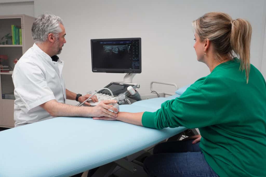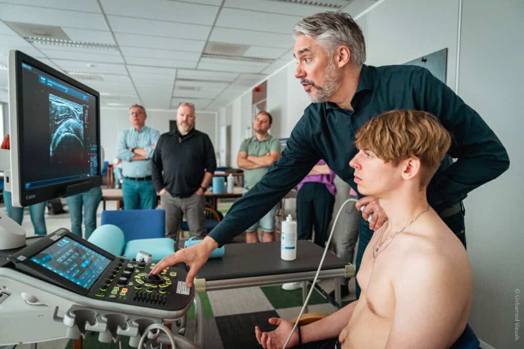
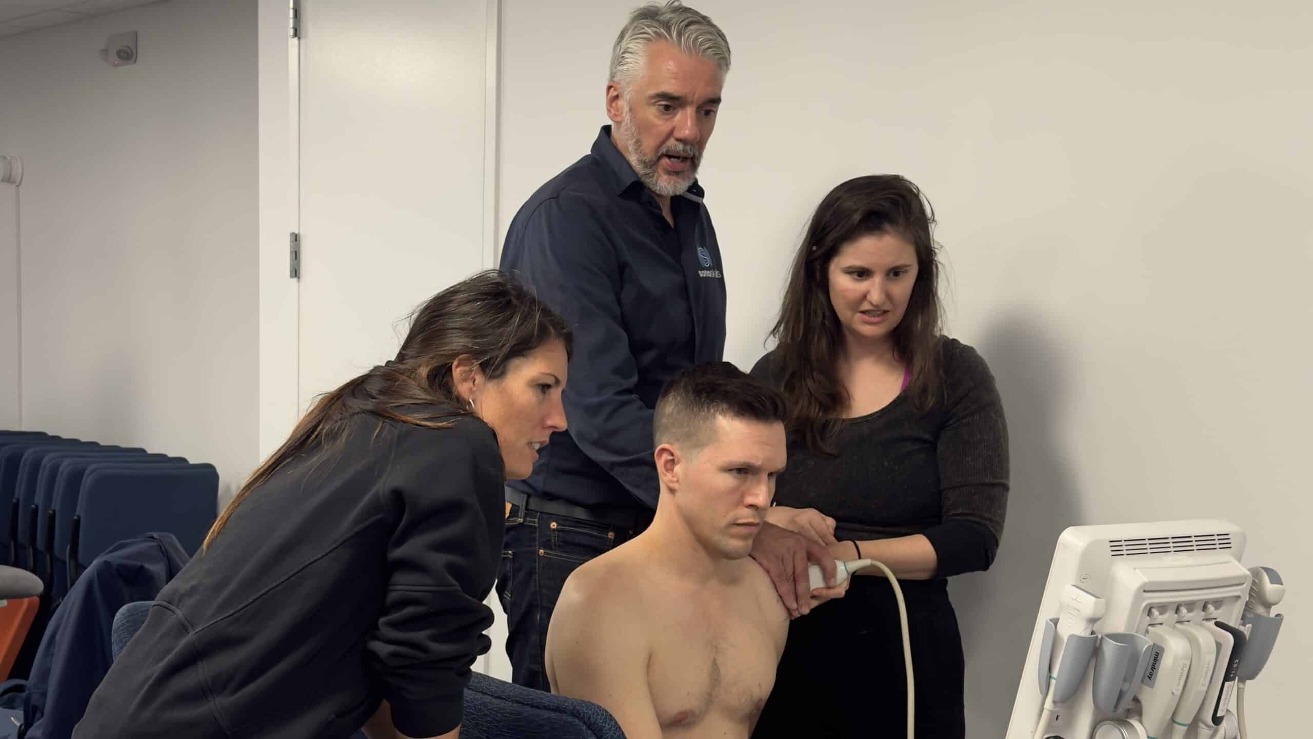
Myopain Seminars is always looking for exceptional companies that offer courses to substantially improve the practice of physical therapy and other healthcare disciplines. In October 2025, Jan Dommerholt contacted SonoSkills, a Dutch company founded by physiotherapist Marc Schmitz, to explore a potential collaboration. Marc and SonoSkills focus on teaching clinicians everything they need to know about Musculoskeletal (MSK) Ultrasound, which in the United States appears to be falling behind several countries, such as Spain, where most orthopedic physiotherapists use ultrasound routinely.
The American Physical Therapy Association (APTA) maintains that “proficient use of ultrasound imaging should provide consistent, high-quality image acquisition and interpretation at the point of care. This helps clinicians refine differential diagnoses in real time, guide targeted interventions, monitor tissue change, and make better decisions about when to continue, escalate, or redirect care. For rehabilitation professionals, these skills strengthen patient education, support safer and more precise care, and help validate clinical findings through real-time imaging.”
In October 2026, Myopain Seminars offered the first SonoSkills MSK Ultrasound course in Bethesda, MD. The comments of the participants speak for themselves:
“Marc is a very skilled and knowledgeable sonographer and teacher. I came in knowing very little about MSK ultrasound and now I feel more confident in how to use ultrasound in my practice or treatment and diagnosis.”
“I did not think I was able to use an ultrasound machine and read images at all after just a weekend. But I did it. Thank you so much for your teaching and patience. The course is worth every penny. Textbook is so helpful, steps by steps instructions. Pre course videos are great. In person class is hands on the entire weekend. You will leave learning what you couldn’t imagine being able to do in just a short time. I’d highly recommend this course to everyone who works in movement and healthcare industry.” — Dr. Linh Taylor, DTCM, LAC, CMTPT
“Very well done. Appreciate the clinical applications throughout the course.”
“I really enjoyed this course. I think this is a very useful skill and I am happy to have been able to learn from Marc.”
MSK ultrasound has evolved into one of the most useful imaging tools in daily musculoskeletal practice. Over the past decade, the technique has gained widespread acceptance among radiologists, sports physicians, orthopedic surgeons, and increasingly physical therapists. The reasons are clear: ultrasound is accessible, dynamic, and relatively inexpensive. At the same time, modern high-frequency probes allow extremely detailed visualization of superficial musculoskeletal structures.
The European Society of Musculoskeletal Radiology (ESSR) periodically reviews the literature and publishes consensus guidelines on the clinical indications for musculoskeletal ultrasound. These guidelines highlight a wide range of conditions where ultrasound is considered a first-line imaging modality or equivalent to other imaging techniques. For clinicians using ultrasound in practice, the key question is simple: which pathologies can we reliably identify with MSK ultrasound?
Below is a practical overview of ten common conditions that musculoskeletal ultrasound can detect, with strong or moderate evidence supporting their use in clinical practice.
- Rotator cuff tears
Rotator cuff pathology is one of the most common reasons for shoulder imaging. MSK ultrasound detects both full-thickness and partial-thickness rotator cuff tears. Ultrasound can demonstrate tendon discontinuity, retraction, and associated fluid in the subacromial bursa. Numerous studies support ultrasound as a first-line imaging technique for rotator cuff tears, particularly when performed by experienced operators.
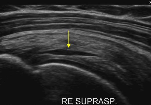
Partial intratendinous supraspinatus tendon rupture in a 77-year old patient with a compaint of shoulder pain.
74-year-old patient presented with severe shoulder dysfunction, weakness, and limited active range of motion. Clinical history likely suggests chronic pathology.
MSK Ultrasound reveals a moderate-to-severe full-thickness tear involving part of the subscapularis tendon width (partial-width), localized to the proximal portion.

- Calcific tendinitis
Calcific tendinitis is frequently seen in the rotator cuff but may also occur in other tendons. On ultrasound, the calcium deposits appear as hyperechoic foci within the tendon, often with acoustic shadowing. One advantage of ultrasound is that it allows dynamic assessment and immediate treatment options such as ultrasound-guided lavage or injections.
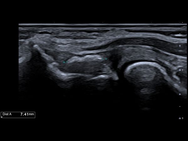
71-year-old male with bilateral lateral elbow pain for more than 18–24 months. Symptoms fluctuate and worsen with loading, gripping, wrist extension, sport, or work-related activity. Previous conservative or interventional treatments have not provided sustained improvement.
MSK Ultrasound showed a tendinopathic change with an intratendinous calcification measuring 7.41 × 2.63 mm in the common extensor tendon (among other findings).
- Lateral epicondylalgia
Lateral epicondylalgia, commonly known as tennis elbow, is a classic indication for MSK ultrasound. Ultrasound typically shows tendon thickening, hypoechoic degeneration, and sometimes Doppler activity reflecting neovascularisation. The literature shows strong evidence supporting ultrasound for evaluating tendon pathology at the lateral epicondyle.
Follow-up ultrasound of the lateral elbow shows persistent enthesophyte or osteophyte formation at the origin of the common extensor tendon.
A previously described partial mid-tendon tear has progressed. It now appears as a full-thickness, partial-width tear of the common extensor tendon. The defect is particularly evident during ultrasound-guided dynamic contraction, where the tendon defect opens, and the full tendon thickness is interrupted across part of the tendon width.
- Carpal tunnel syndrome
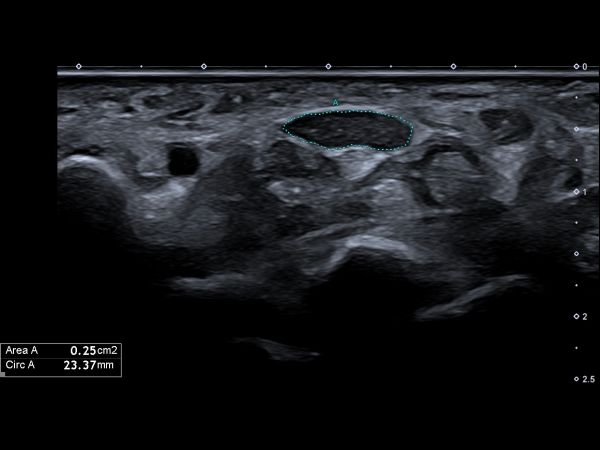
Peripheral nerve imaging is a growing area within musculoskeletal ultrasound. Carpal tunnel syndrome is one of the most widely studied examples. Ultrasound can show median nerve enlargement, changes in nerve echotexture, and structural causes of compression. These findings correlate well with electrodiagnostic testing and can support the clinical diagnosis.
49-year-old patient presented with symptoms suggestive of peripheral nerve compression, including possible numbness, tingling, or weakness in the hand and forearm.
At the level of the carpal tunnel, the median nerve is markedly enlarged and hypoechoic, consistent with nerve edema and structural alteration. The cross-sectional area measures approximately 25 mm², which is clearly above the commonly accepted pathological threshold of approximately 12 mm². These findings are consistent with carpal tunnel syndrome.
- De Quervain’s tenosynovitis
De Quervain’s disease affects the tendons of the first dorsal compartment of the wrist. Ultrasound typically shows tendon sheath thickening and fluid surrounding the tendons. Because of its high spatial resolution, ultrasound is particularly well suited for detecting tenosynovitis in small tendon compartments of the wrist and hand.
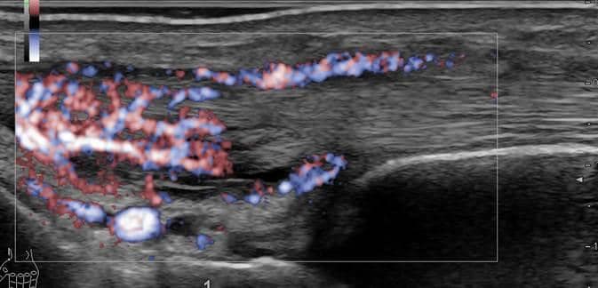
50-year-old female with swelling on the dorsal radial side of the wrist.
MSK Ultrasound showed De Quervain’s tenosynovitis with a thickened, hypervascularized tendon and synovium.
- Baker’s cyst
A Baker’s cyst is a fluid collection in the popliteal fossa, typically arising between the semimembranosus tendon and the medial head of the gastrocnemius muscle. Ultrasound easily detects these cysts and helps differentiate them from other masses in the popliteal region. Ultrasound can also guide aspiration if treatment is required.
Fluid distension of the semimembranosus–gastrocnemius bursa is present, compatible with a Baker cyst.
- Joint effusion and synovitis
Ultrasound is very sensitive for detecting fluid within joints. Ultrasound can visualize small joint effusions in the shoulder, elbow, knee, ankle, or wrist. In inflammatory conditions, ultrasound can also detect synovial hypertrophy and increased vascularity using Doppler imaging. This makes ultrasound useful not only for diagnosis but also for monitoring inflammatory joint diseases.
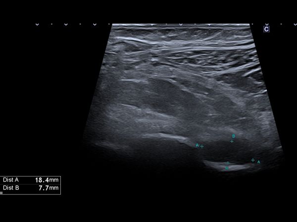
59-year-old patient presented with hip-related complaints.
Ultrasound examination demonstrates mild irregularity of the superficially visible anterior cortical contours of the hip joint, most likely degenerative in origin.
Additional irregularities are present at the level of the femoral neck (collum femoris) and femoral head (caput femoris), also suggestive of degenerative osseous remodeling.
There is increased fluid within the hip joint capsule, corresponding to grade 3 of 3 on the synovitis scoring system, indicating marked capsular distension / active synovitis.
- Achilles tendinopathy
Achilles tendinopathy is another classic musculoskeletal ultrasound application. Typical findings include tendon thickening, loss of normal fibrillar structure, and sometimes intratendinous Doppler signal. Ultrasound is particularly valuable because it allows dynamic examination and easy comparison with the contralateral side.
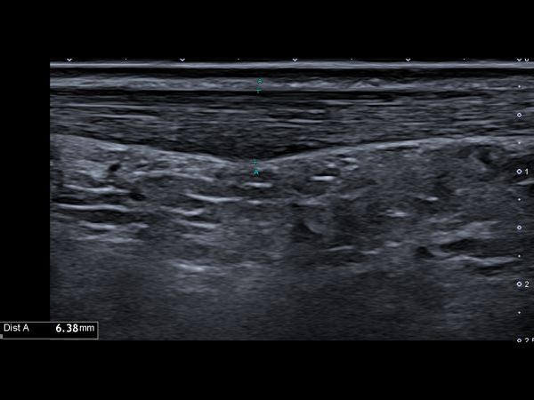
20-year-old patient with right ankle and/or Achilles region symptoms. Ultrasound examination was performed of the Achilles tendon, flexor hallucis longus, tibialis posterior, flexor digitorum longus, fibularis longus and brevis, talocrural joint, retrocalcaneal bursa, medial and lateral ankle ligaments, and syndesmosis.
The Achilles tendon shows moderate mid-portion tendinopathy. No partial or complete Achilles tendon rupture is present.
- Plantar fasciitis
Plantar fasciitis is one of the most common causes of heel pain. On ultrasound, the plantar fascia typically appears thickened and hypoechoic near its calcaneal insertion. In some cases, Doppler signal may indicate active inflammation. Ultrasound is widely used to confirm the diagnosis and to guide therapeutic injections.
39-year-old female with heel pain presented with a hypoechoic thickened plantar fascial insertion.

- Morton’s neuroma
Morton’s neuroma is a benign enlargement of the plantar digital nerve usually located between the third and fourth metatarsal heads. Ultrasound can demonstrate a hypoechoic mass in the intermetatarsal space. In experienced hands, ultrasound has good diagnostic accuracy and can also guide corticosteroid or alcohol injections.
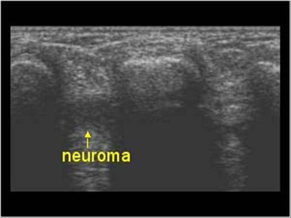
Mortons neuroma with a hypoechoic mass in the interdigital space and fluid bursa.
In summary, musculoskeletal ultrasound has become an essential imaging technique for evaluating many common musculoskeletal disorders. It is particularly well suited for examining superficial structures such as tendons, ligaments, bursae, and peripheral nerves. The ESSR consensus guidelines confirm that ultrasound plays a major role in assessing degenerative and traumatic tendon disorders, as well as several other soft tissue conditions.
For clinicians who integrate ultrasound into their daily practice, the technique offers an important advantage: it combines imaging, clinical examination and interventional procedures in a single session. With appropriate training and experience, MSK ultrasound becomes a powerful extension of the physical examination and a valuable tool in modern musculoskeletal care.
Jan Dommerholt, PT, DPT, President / CEO, Myopain Seminars
You can find the images and case reports in the free ultrasound library offered by SonoSkills. Inspired by SonoSkills Articles.