
Jan Dommerholt, PT, DPT | President/CEO, Myopain Seminars
Introduction
When Robert Gerwin, MD, and Jan Dommerholt, PT, DPT, started teaching the first dry needling courses in Spain (1996) and the United States (1997), we focused almost exclusively on dry needling of trigger points. Our dry needling “brand” evolved from the practice of trigger point injections developed by our mentor, Dr. Janet Travell. Although Travell mentioned dry needling in a few of her articles, she used the term when she used a hypodermic needle but did not inject any fluid, hence the term “dry” needling. Dr. Travell was the White House physician of Presidents John F. Kennedy and Lyndon B. Johnson. Bob Gerwin worked with Travell for many years, while Jan Dommerholt met her during a 1989 workshop she taught at the National Rehabilitation Hospital in Washington, DC. Dr. Travell was 89 years old.
Several years later, Bob and Jan started the Janet G. Travell, MD Seminar SeriesTM in honor of her contributions to the field of myofascial pain. Eventually, we changed the name of our course program to Myopain Seminars.
Following our initial focus on trigger points, several other dry needling targets have evolved, such as scar and fascial needling, periosteal needling, needling for recovery, needling to reduce spasticity, tendon and enthesal needling, and perineural needling.
Diabetic Foot Ulcers
One of the more intriguing applications of dry needling was investigated by Fatemeh HasanNia from the Department of Physiotherapy, School of Rehabilitation, Tehran University of Medical Sciences in Iran. Two patients with neuropathic diabetic foot ulcers, who were unresponsive to conventional wound care, received eight dry needling sessions. Traditional treatments may include surgical debridement, antibiotics, vascular assessment, offloading, and even amputation, but unfortunately, these methods are often inadequate and result in prolonged healing times. There was some preliminary evidence in favor of dry needling for idiopathic peripheral neuropathy (Nasr and Zafereo, 2019), but no one had considered using dry needling to treat diabetic foot ulcers. I was a member of her advisory team, and frankly, I was skeptical about conducting this case study, especially when the student proposed to only needle select calf muscles!
The first patient was a 56-year-old male patient with a 20-year history of diabetes who presented with a persistent neuropathic foot ulcer on the plantar aspect of the right fifth metatarsal, classified as a grade 2 ulcer according to the Wagner Classification System (Oyibo, et al., 2001). Traditional treatments, including dressing changes, debridement, and glycemic control, did not alter the ulcer. A 0.3 x 50 mm needle was used to treat the medial and lateral head of the gastrocnemius, the distal medial and lateral soleus, flexor hallucis longus, flexor digitorum longus, tibialis anterior, fibularis longus, and extensor hallucis longus muscles. After eight dry needling sessions over four weeks, consisting of the fast in-out technique and leaving the needles in place for ten minutes, a follow-up assessment was scheduled two months after the last treatment session. Surprisingly, the ulcer area was reduced by 65% and entirely healed within two months.
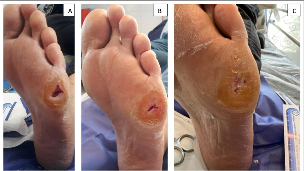
The ulcer condition in the 1st week (A), 4th week (B), and 12th week (C).
The second case was a 56-year-old woman suffering from an ulcer for the past 7 months that was also unresponsive to traditional treatments. The treatment protocol was the same as for the first case, but the plantar ulcer healed after eight sessions of dry needling in this case.
The ulcer condition in the 1st week (A) and 4th week (B)
In the paper (HasanNia et al., 2024), which is available at the Myopain Seminars website, we only speculated about the possible underlying mechanisms of wound healing following dry needling and suggested that central sensitization and myofascial dysfunction could have contributed to the neuropathy and delayed ulcer healing. However, more research and clinical studies are needed.
Fascia and Wound Healing
A recent paper by Xu and colleagues (2025) explored the possible role of fascia in skin wound healing. The authors emphasized that fascia not only provides mechanical support but also contributes to tissue repair and intercellular signaling. Fascia is rarely considered in descriptions of the mechanisms of wound healing, as the focus is typically on the repair mechanisms of the epidermis and dermis. The authors suggested that, especially in chronic wound healing, changes in the fascia may affect long-term wound repair, leading to healing complications and disorders.
In patients with diabetes, tissue oxygenation and oxygen transport may be impaired because of hyperglycaemia, endothelial dysfunction, and a decrease in microcirculation, which will negatively impact the healing process, leading to peripheral neuropathy and vascular disease. Furthermore, the local inflammatory response may be impaired due to an impaired immune response, macrophage and neutrophil activity, which may increase the risk of infection.
Correa-Gallegos and colleagues (2022) suggested that diabetic and ulcerative wounds, as well as hypertrophic scars, may be attributed to the fascia and, in particular, to fibroblast activity. Xu and colleagues noted, “Fascia is not only a structural bridge connecting the skin to deeper tissues but also a crucial regulatory factor in wound repair.” A detailed review of the mechanisms is beyond the scope of this blog, but we have included the paper as a download.
In summary, we are optimistic that other practical applications of dry needling will be discovered. For diabetic neuropathic ulcers, a better understanding of the potential role of fascia in wound healing may lead to developing more effective individualized and fascia-targeted treatment strategies.
References
Nasr, A. J., & Zafereo, J. (2019). The effects of dry needling and neurodynamic exercise on idiopathic peripheral neuropathy: A case report. Journal of Bodywork and Movement Therapies, 23(2), 306–310. https://doi.org/10.1016/j.jbmt.2018.02.006
Oyibo, S. O., Jude, E. B., Tarawneh, I., Nguyen, H. C., Harkless, L. B., & Boulton, A. J. (2001). A comparison of two diabetic foot ulcer classification systems: the Wagner and the University of Texas wound classification systems. Diabetes Care, 24(1), 84–88. https://doi.org/10.2337/diacare.24.1.84
HasanNia, F., Amini, M. R., Sanjari, M., Ansari, N. N., Dommerholt, J., Khalifelou, M., & Naghdi, S. (2024). The effect of dry needling on the healing process of neuropathic diabetic foot ulcers: case study of two patients. Case Rep Orthop Surg J, 3(4), 143.
Xu, J., Zhang, H., & Ye, H. (2025). Research progress on the role of fascia in skin wound healing. Burns Trauma, 13, tkaf002. doi:10.1093/burnst/tkaf002
Correa-Gallegos, D., Jiang, D., Christ, S., Ramesh, P., Ye, H., Wannemacher, J., . . . Rinkevich, Y. (2019). Patch repair of deep wounds by mobilized fascia. Nature, 576(7786), 287–292. doi:10.1038/s41586-019-1794-y