Introduction: Persistent Arthrogenic Muscle Inhibition After Knee Injury
Recovery following knee ligament injury is a multifactorial process that extends well beyond tissue-healing timelines. While ligament integrity, joint stability, and pain resolution are critical milestones, neuromuscular recovery frequently lags behind structural repair. One of the most significant contributors to this delayed functional recovery is arthrogenic muscle inhibition (AMI)—a reflexive reduction in muscle activation driven by altered sensory input from an injured joint.
AMI is particularly impactful in the quadriceps muscle following knee injury or surgery. Altered afferent input from pain, swelling, inflammation, and joint mechanoreceptor disruption leads to reflex inhibition at both spinal and supraspinal levels, significantly reducing voluntary activation of the quadriceps [1]. Importantly, even small joint effusions or minimal pain can produce substantial inhibition, independent of measurable strength loss.
The clinical relevance of AMI lies in its persistence. Quadriceps inhibition may persist long after swelling has resolved and strength appears to be restored, contributing to a delayed return to sport, altered movement strategies, and elevated risk of reinjury. However, joint-driven AMI represents only one component of post-injury neuromuscular dysfunction. Psychological factors such as fear, confidence, and motor relearning play a role, as do non-articular tissue influences, including inhibitory mechanisms originating within the muscle itself.
Clinical Background and Functional Deficits
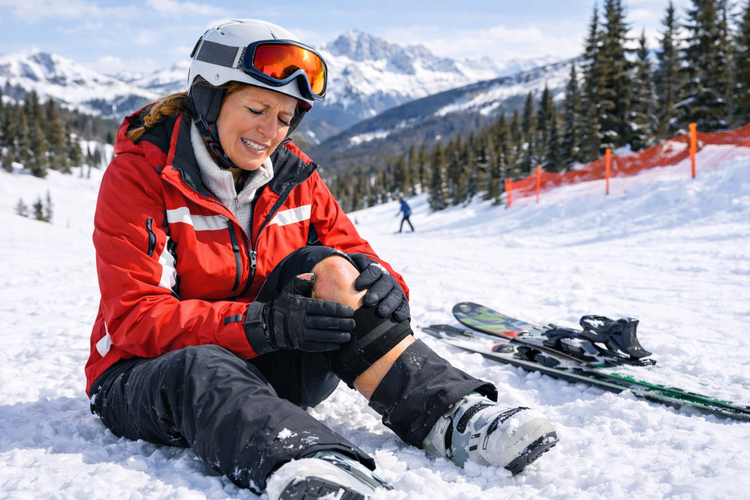
The patient was a 46-year-old female alpine skier who sustained partial tears of the medial collateral ligament (MCL) and anterior cruciate ligament (ACL) of her left knee on February 22, 2025. By August, she had progressed through rehabilitation with restoration of joint range of motion and reported no resting knee pain or instability.
Hand-held dynamometry demonstrated quadriceps and hamstring strength within 10% of the uninvolved limb, a symmetry commonly considered acceptable for return to activity. However, deeper neuromuscular assessment revealed persistent deficits. Surface electromyography (sEMG) demonstrated a 32% deficit in peak voluntary quadriceps contraction on the injured side compared to the contralateral limb.
The clinical significance of these findings was reinforced by functional testing. Single-leg jump height on the injured limb was only 67% of that on the uninvolved side, and the patient continued to demonstrate difficulty with eccentric quadriceps control, particularly during step-down tasks, stair descent, and plyometric activities. Despite continued strength training, progress in these domains had plateaued.
For an alpine skier—where controlled eccentric loading, rapid force modulation, and dynamic knee stability are paramount—these deficits represented a meaningful barrier to performance and safe return to sport.
Beyond the Joint: Intramuscular Inhibition and Myofascial Trigger Points
While AMI is classically attributed to altered joint afferent input, emerging evidence supports the role of intramuscular inhibitory mechanisms that may persist independently of joint pathology. Myofascial trigger points (TrPs) represent one such mechanism.
Trigger points are localized, hyperirritable regions within skeletal muscle associated with abnormal endplate activity and altered motor control. Importantly, latent trigger points, although not producing spontaneous pain, impair muscle activation, force production, and coordination [2]. This distinction is clinically critical, as the absence of pain does not equate to normal muscle function.
Dry needling of myofascial trigger points can improve muscle activation, EMG amplitude, and neuromuscular performance across multiple muscle groups, including the quadriceps [2]. These effects are attributed to normalization of motor endplate efficiency, reduction of inhibitory afferent input, and improved corticospinal drive.
Given the patient’s persistent activation deficits despite normalized strength and absence of joint symptoms, intramuscular inhibition was considered a plausible contributor to her ongoing dysfunction.
Intervention: Dry Needling of Latent Trigger Points in the Vastus Lateralis
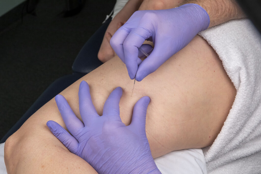
Although the patient did not report quadriceps pain, palpation identified latent myofascial trigger points within the vastus lateralis of the injured limb. A single session of myofascial trigger point dry needling was performed, with needle placement directed to elicit a local twitch response (LTR).
The importance of achieving an LTR is well supported in the literature. Local twitch responses reflect a spinal reflex mediated by abnormal endplate activity. They are associated with greater reductions in spontaneous electrical activity and improved muscle function compared to needling without twitch elicitation [3,4]. Studies by Hong and others have demonstrated superior outcomes in muscle activation and force production when LTRs are achieved [3].
Outcomes: Immediate and Short-Term Improvements in Neuromuscular Function
The response to the intervention was clinically meaningful. Immediately post-treatment, sEMG assessment demonstrated improved peak quadriceps activation, reducing the deficit from 32% to 14% relative to the uninvolved limb. Concurrently, the patient showed a notable improvement in step-down performance, with improved speed control and elimination of the previously observed loss of eccentric control during terminal descent.
Within the following two weeks, single-leg jump height improved to within 10% of the uninvolved side, representing a substantial functional gain. These changes occurred without alterations in strength training volume or loading parameters, supporting a neuromuscular rather than hypertrophic mechanism of improvement.
Mechanisms: How Trigger Points Inhibit Muscle Force Production
Trigger points interfere with muscle contraction through multiple mechanisms. Sustained sarcomere shortening within taut bands reduces effective force transmission, while abnormal endplate noise increases metabolic demand and disrupts motor unit recruitment [4]. Additionally, both nociceptive and non-nociceptive afferent input from trigger points may contribute to central inhibition, further reducing voluntary activation—even in latent, non-painful states [2].
Dry needling appears to reverse these effects through mechanical disruption of dysfunctional motor endplates, normalization of local biochemical milieu, and reduction of inhibitory afferent input. Some evidence suggests that these effects are enhanced when a local twitch response is elicited, supporting the clinical emphasis on twitch-responsive needling techniques [3,4].
Conclusion
This case highlights the importance of comprehensive neuromuscular assessment following knee ligament injury. While arthrogenic muscle inhibition remains a well-established contributor to quadriceps dysfunction, intramuscular inhibitory mechanisms such as latent myofascial trigger points may independently perpetuate activation deficits, even when strength appears normalized.
In this alpine skier, a single session of trigger point dry needling to the vastus lateralis—despite the absence of pain—was associated with immediate improvements in quadriceps activation and meaningful short-term gains in functional performance. These findings support integrating muscle-based interventions with joint-focused rehabilitation strategies to address persistent neuromuscular inhibition and return-to-sport limitations.
Ralph Simpson, PT, DPT, OCS, CMPT, CMTPT, LATC – Instructor
References
- Rice DA, McNair PJ. Quadriceps arthrogenic muscle inhibition: neural mechanisms and treatment perspectives. Sports Med. 2010.
- Gattie E, Cleland JA, Snodgrass S. The effectiveness of trigger point dry needling for musculoskeletal conditions: a systematic review. J Orthop Sports Phys Ther. 2017.
- Hong CZ. Lidocaine injection versus dry needling to myofascial trigger point: the importance of the local twitch response. Am J Phys Med Rehabil. 1994.
- Shah JP et al. Biochemical milieu of myofascial trigger points before and after dry needling. Arch Phys Med Rehabil. 2008.
- Hermens HJ et al. SENIAM recommendations for surface electromyography. 1999.